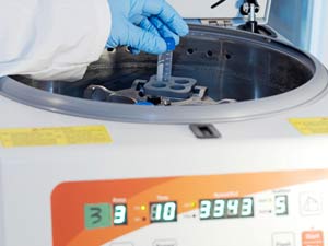
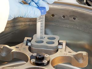
1. Blood samples are loaded into the centrifuge to extract tumor-specific exosomic DNA for our proprietary process.
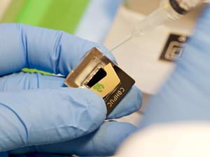
2. Once the DNA is extracted, it is loaded onto the QuantStudio 3D V2 chips. The samples are loaded using the latest in digital bluetooth micro pipettes.
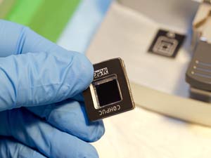
3. Each chip contains 20,000 individual wells. The tumor DNA is bound to the FAM probe, and the wild type (normal version of the DNA) is bound to the VIC probe. Each well contains either a VIC or FAM labeled piece of DNA
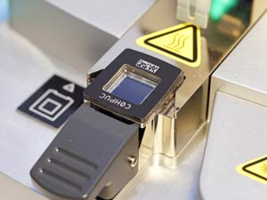
4. The chip is then sealed using ultraviolet light.
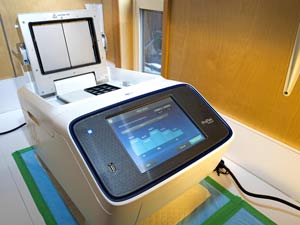
5. The chips are then loaded into a flat block thermal cycler and the DNA is amplified.
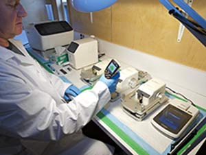
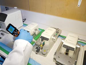
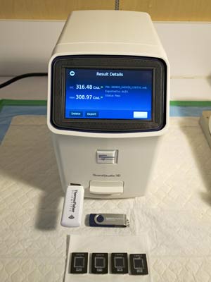
6. The chips are then read by the chip-reader, which contains a high powered laser. The laser photographs each individual well, and determines the ratio of the FAM to the VIC.
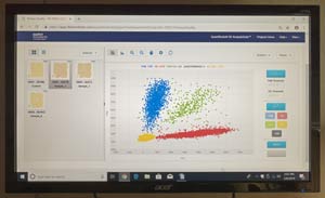
7. The chip reader automatically uploads the data to the online software analysis cloud and analyzes it. This picture shows the FAM (in blue) and the VIC (in red). The ratio of the FAM and the VIC gives us the percentage needed for the final report.